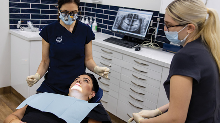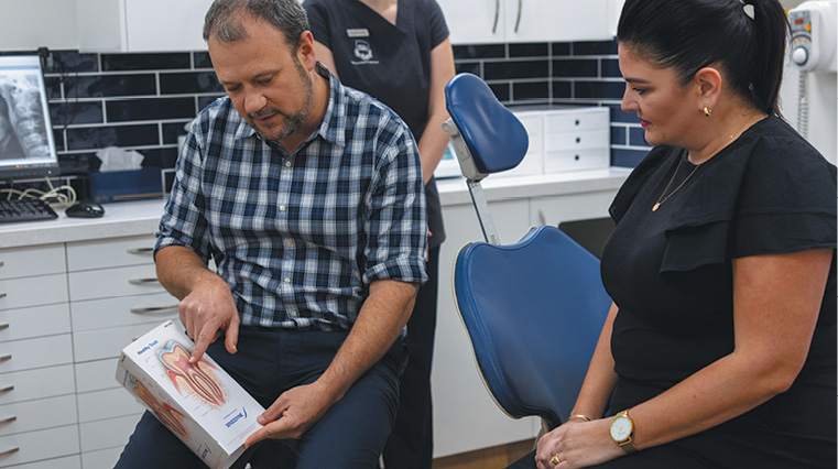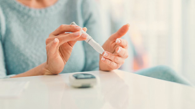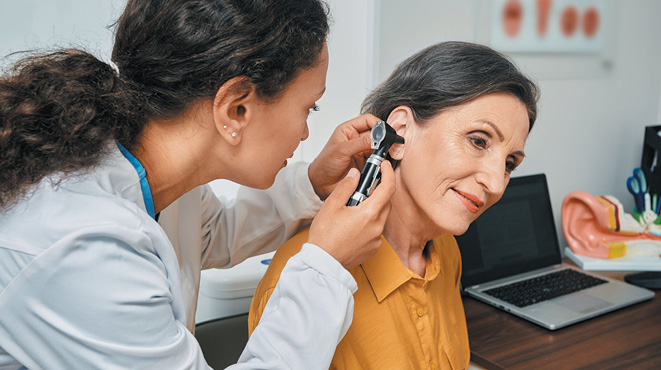
BY DR ROGER DOWN, MBBS, FRCS, FRACS, MD (LOND)
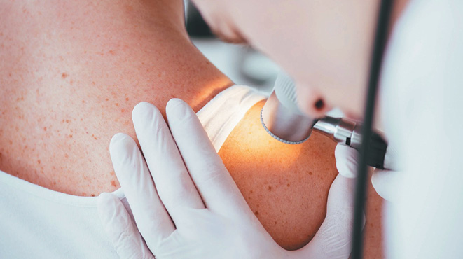
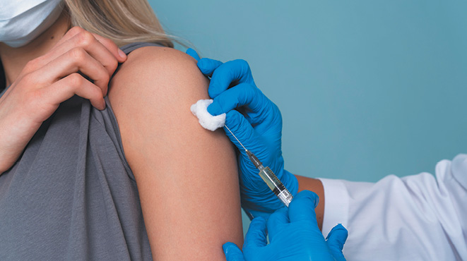
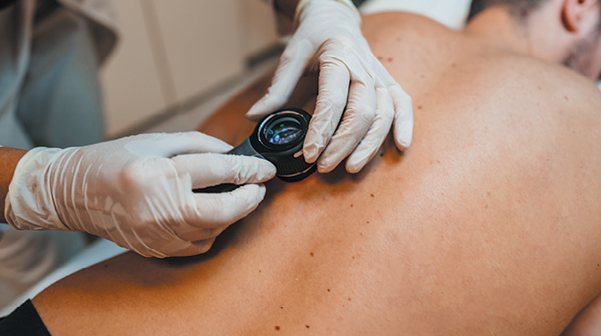
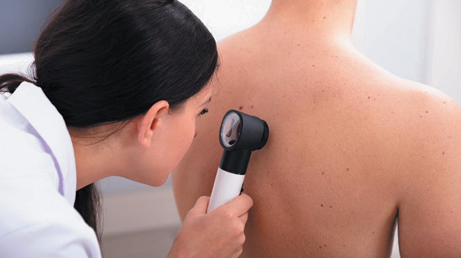
 All surgically removed tissue must be sent for histopathology to determine the nature of the lesion. This is why I do not advocate freezing, burning, or chemotherapeutic agents like Efudix, as no pathology specimen is obtained. There is a view that cryotherapy can do ‘no harm’. This is not true. It opens Pandora’s Box, and is subject to over-treatment for reward.
Histopathology can determine whether a lesion is benign or malignant and identify the cancer cell type. Firstly, this is important for determining the patient’s likely prognosis and guiding the specialist’s treatment. Secondly, it can provide the clinician with guidance on whether all the cancer has been removed.
Please remember that the skin cancer cure is related to the tissue/cells that remain in the patient, not to what the pathologist sees. The pathologist’s report is based on random sections, which are subject to contraction, folding, and deformity, which can, in turn, provide misleading information. Further, fat gets dissolved, often leading to an incorrect report, which reads, ‘extends to the deep margin’.
So, what matters is as follows: are there any residual cancer cells in the patient that were not removed? This is impossible to say for certain. Single or multiple cancer cells can spread via the lymphatics or the bloodstream. A pathology report cannot always determine this, but it is looked for.
Dogma dictates that ‘margins’ (i.e., the distance from the cut edge to the cancer edge) determine this, but experience with 100,000s of cases and studies negates this view. All tumours must be macroscopically removed, with a small additional ‘normal’ tissue margin. The surgeon’s competence matters.
The pathologist is looking at slides but cannot determine this with certainty; they can only state what is seen.
Personal studies of thousands of patients show that 90-95% of ‘margin involved’ pathology reports – especially after a partial-thickness skin shave excision of intraepidermal cancers – were followed up three-monthly over two years and never developed a recurrence. Of those patients who did have an early second re-operation because of site complexities, the pathologists were unable to find any residual cancer in 95% of the specimens. This leads to the conclusion that ‘out is out’. With the exception of melanomas, the margin is most likely irrelevant.
Are the very small number of patients with ‘margin involved’ pathology reports in danger? Basically, no.
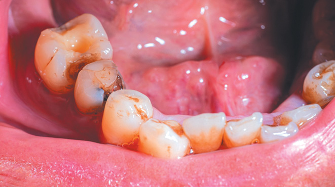
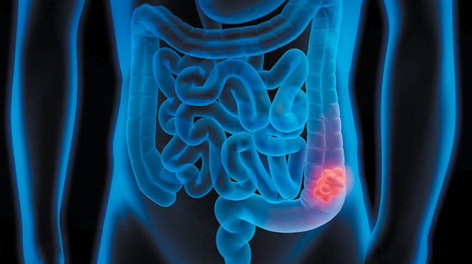
Basal cell cancers only grow locally and do not spread or metastasise to other parts of the body. About 10% of invasive squamous cell cancers (they must always have a 1-2mm clear margin in all planes) that penetrate the full skin thickness can metastasise or develop a local recurrence. Local recurrence needs to be re-excised as soon as it declares itself, usually without detriment. Very rarely, the ‘horse may have bolted’ prior to the original surgery, leading to the development of lymph node metastasis, which can then easily be treated with either surgery or DXT.